")

Flow Cytometry Services
About the service
Flow cytometry technique is a powerful method for counting and analyzing fluorescently stained cells in a single cell population to identify the presence of antigen molecules on the surface or inside the cell, thereby facilitating the interpretation of cellular events, including processes, conditions, and structures associated with cellular function and physiology. In this technique, cells are labeled with fluorochromes, allowing for their identification and measurement in two distinct modes: indirect detection using antibodies or direct detection without fluorochrome binding to the cell.
Important applications of flow cytometry technique
- Immunophenotyping: By employing antibodies conjugated with fluorescence substances for specific antigen staining, this technique enables the identification of the percentage of positive cells and the relative amount of antigen expression within the cell population.
- Apoptosis: The rate and percentage of apoptosis and necrosis are determined with the help of Annexin-PI staining among the cell population.
- Cell Cycle Analysis: This test examines the amount of genetic material within cells and determines the phase of the cell cycle (G2, S, G1, and mitosis) in which the cells within the population reside. The chromatin content of the cells is stained using PI or DAPI, allowing for accurate analysis.
- Cell Cycle Analysis and Immunophenotyping: This technique enables the examination of the expression level of the target antigen within the cell population and explores the relationship between antigen expression and the cell division cycle, including its phases.
- Viability Assessment: The viability and death rate of cells within the population can be determined by employing fluorochromes that can bind to the genetic material, cell membrane, or free amines present on the cell surface and inside the cell. This technique enables the assessment of cell viability within the analyzed cell population.
- Cell Proliferation Analysis: The flow cytometry technique, combined with CFSE staining, allows for convenient assessment of the cell proliferation rate across various generations.
- Cell Size and Granularity Analysis: One of the fundamental functionalities of flow cytometry is the capacity to assess and display the relative size and granularity of cells.
Basic and technical concepts in using flow cytometer
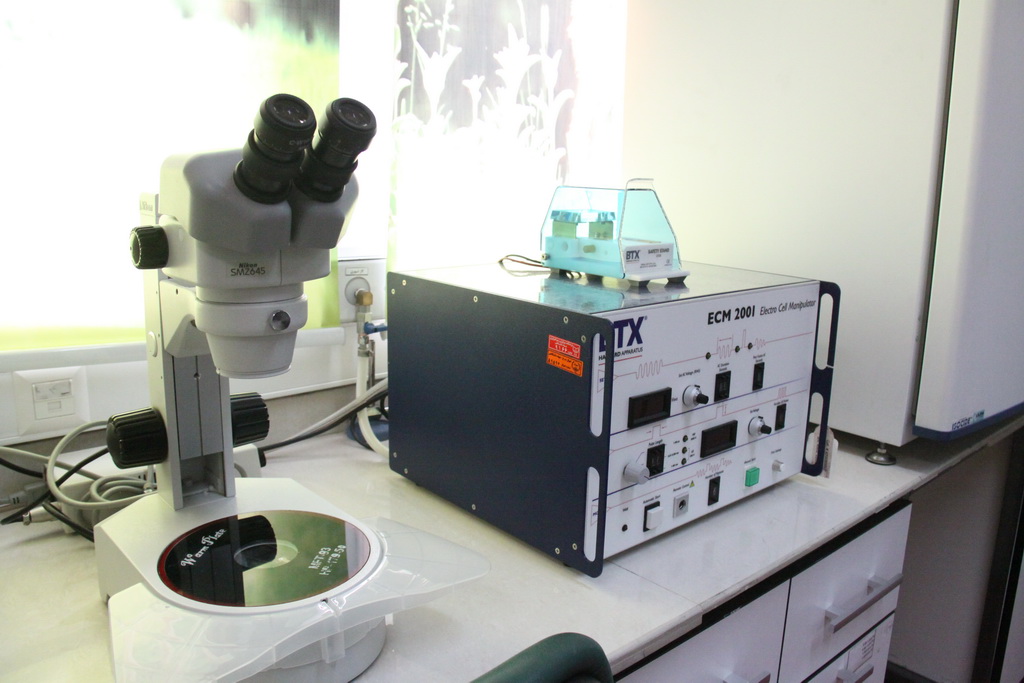
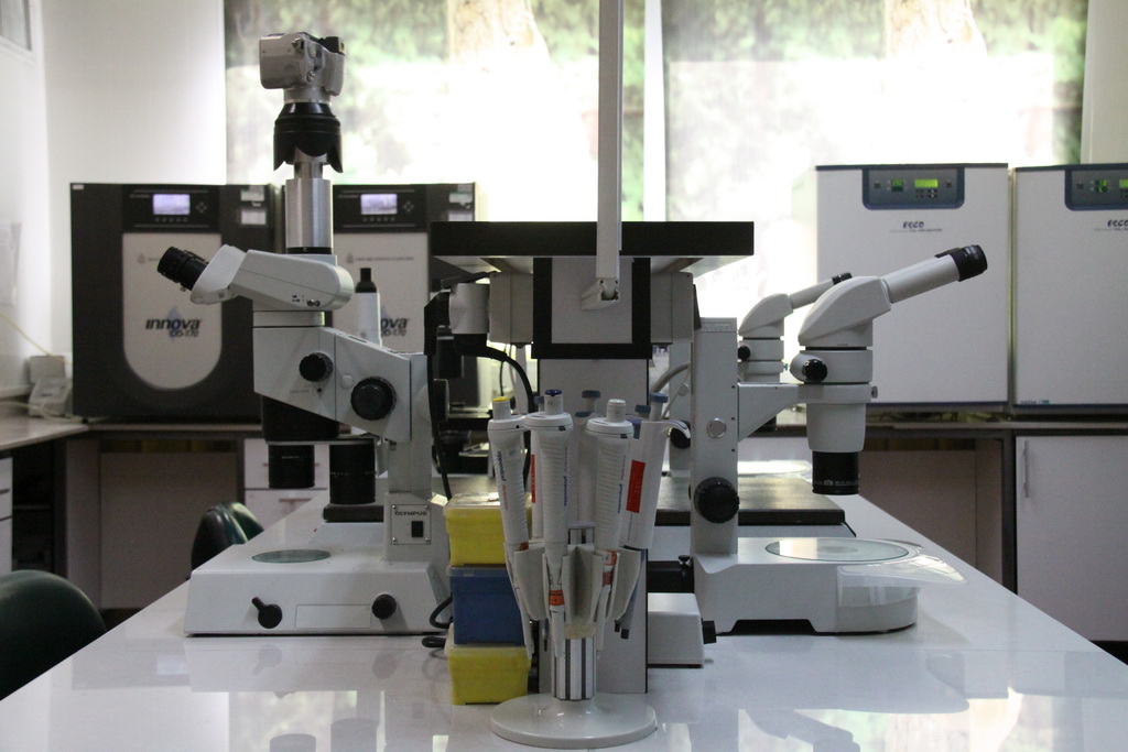
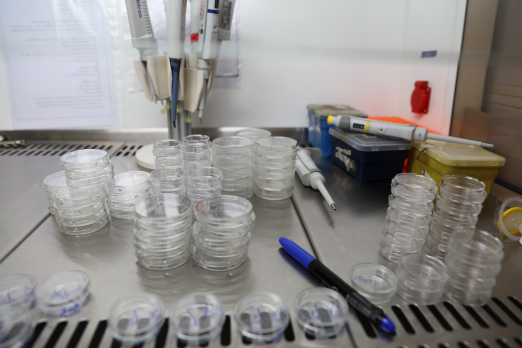
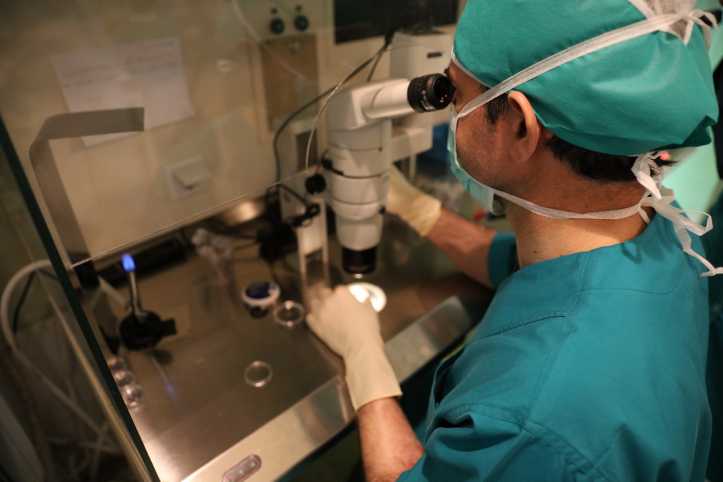
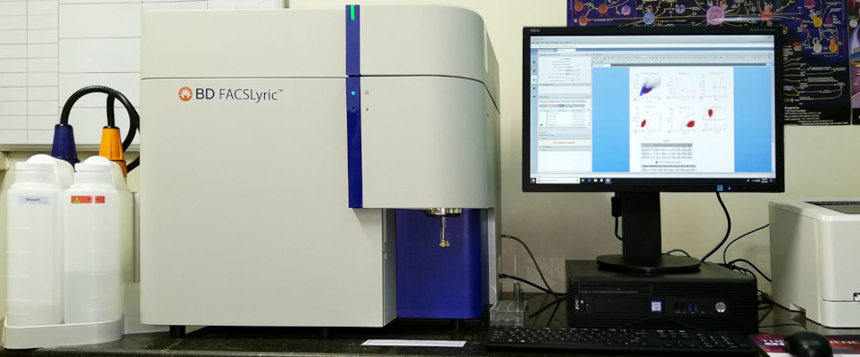
To conduct flow cytometry, individual cells need to be collected and suspended in an appropriate medium. Once a suspension containing a sufficient number of at least 200,000 cells is prepared, the cells are labeled with fluorochrome-conjugated antibodies following the desired protocol. After washing and centrifugation steps, the labeled cells are transferred to the flow cytometry laboratory in an albumin-containing buffer (0.5%) on ice, while ensuring protection from light. Once resuspended, the cells are introduced into the flow cytometer, where they are counted and recorded based on the type and intensity of the fluorescence signal. This laboratory is equipped with a FACSLyric flow cytometer and two PARTEC PAS flow cytometers.
Flow cytometry equipment
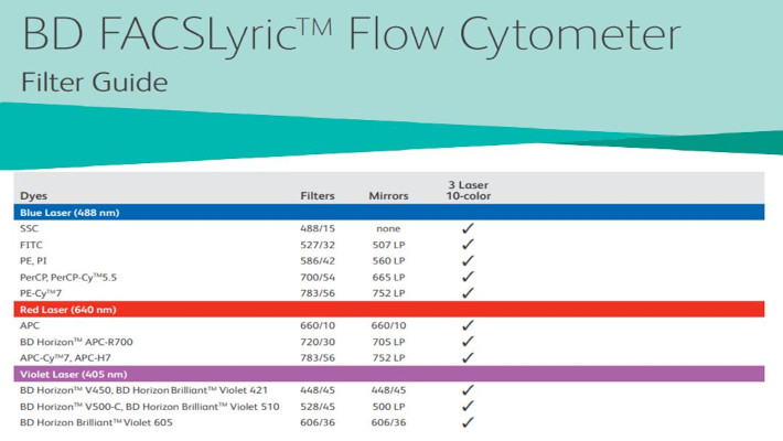
- BD FACSLyric flow cytometry device is equipped with three 488, 640 and 405 nm lasers. The details of lasers and the filters can be seen in the table below.
- The laboratory is equipped with two PARTEC PAS-II flow cytometers, each featuring two 488 nm argon ion laser sources, a 635 nm diode laser, and an HBO lamp for UV radiation

Laboratory Supervisor:
Laboratory Assistant:
Zhamak Pahlevanzadeh (MSc in Cellular and Molecular Biology); Email: ; Contact Number: +98 (21) 22432020 - Extension 205
Location of Flow Cytometry Laboratory:
The laboratory is situated on the first floor of Avicenna Research Institute, located within Shahid Beheshti University.
Working Schedule:
The laboratory operates from Saturday to Wednesday, between 8:00 AM and 4:00 PM.
Service Policy and Costs:
The cost of tests or services will vary depending on the sample type and specific testing requirements. Therefore, upon the initial inquiry, the laboratory will provide information regarding the cost of the requested test.
Sample Submission:
To ensure smooth processing, please coordinate with the laboratory assistant at least 48 hours before sending the sample.
Further Resources
- BD TRAINING & E-LEARNING: www.bdbiosciences.com/eu/s/training/e-learning
- BD Spectrum Viewer: www.bdbiosciences.com/en-us/applications/research-applications/multicolor-flow-cytometry/product-selection-tools/spectrum-viewer
- Fluorescence Tutorials: www.thermofisher.com/nl/en/home/support/tutorials.html
- Thermo Fluorescence SpectraViewer: www.thermofisher.com/nl/en/home/life-science/cell-analysis/labeling-chemistry/fluorescence-spectraviewer.html