Light Microscopy–Fluorescence Services
About the service
Avicenna Research Institute's light-fluorescence microscope laboratory is equipped with both upright and inverted light-fluorescence microscopes, enabling researchers and students to observe and capture images of cell and tissue samples on standard slides or culture media. Since cell and tissue samples are transparent and lack sufficient contrast, the observed image may not reveal distinguishable or reliable details. To address this, various methods have been developed to color cell or tissue components and enhance contrast, thereby displaying sample details. It is important to note that the choice of staining protocol for cells or tissues determines whether the observation should be conducted using the light microscope mode or the fluorescence microscope mode. When chemical-chromogenic methods (cytochemical and histochemical methods) are employed to stain samples, the optical part of these microscopes can be used for capturing photographs. On the other hand, samples stained using fluorochromes and following immunofluorescence protocols require the fluorescence capabilities of the microscope for observation and photography.
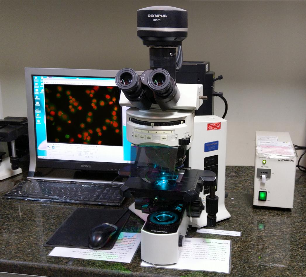
1. The Olympus BX51 upright microscope is equipped with a DP71 highly sensitive camera. The Olympus BX51 upright microscope, featuring a DP71 highly sensitive camera, requires samples to be prepared on glass slides for observation and imaging.
- For samples that have been chemically stained and mounted using Entellan® adhesive, they can be observed and photographed using the optical mode of the microscope. These slides do not necessitate any specific precautions or additional care like fluorescence samples.
- In the case of fluorescence samples prepared on glass slides, a mounting solution of PBS-Glycerol in a 50/50 ratio is typically used. To prevent the slides from drying out, they should be placed in light-impermeable plastic boxes with a small amount of water at the bottom. These slides can be stored in a cold environment at temperatures between 4 and 8 degrees Celsius for up to one week, ensuring proper moisture retention.
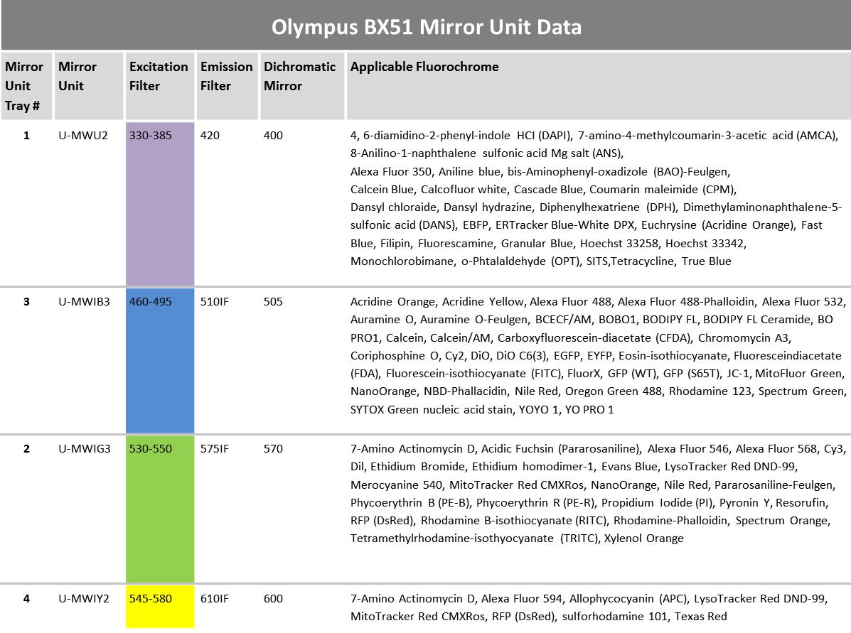
- Regarding the fluorescence capabilities of the microscope, the light emitted by the mercury lamp of the microscope is used in conjunction with filters that have average wavelengths of 366, 477, 546, and 579 nm. These specific wavelengths can be employed for excitation and to facilitate the visualization of desired fluorochromes. The microscope's lenses offer magnifications of ×4, ×10, ×20, ×40, and ×100.
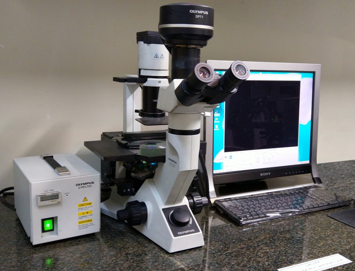
2. The Olympus CKX41 inverted microscope, equipped with a highly sensitive DP71 camera, allows for the observation and photography of cell samples within cell culture plates or flasks using both the optical and fluorescence sections. The microscope lenses can be utilized at three magnifications of ×4, ×10, and ×40. Additionally, this microscope is equipped with two filters with average wavelengths of 477 nm and 546 nm for the excitation of desired fluorochromes. This laboratory has a long and successful history in photographing animal and plant cell samples, as well as transparent synthetic samples, with a maximum resolution of 500 nm.
Below are a series of photographs captured using an Olympus-BX51 upright microscope:
- Slide prepared using the Cytospin method, stained with FITC and DAPI, and observed through two filters: 460-495 nm (FITC labeling the cell membrane) and 330-385 nm (DAPI highlighting the nucleus)
- Slide prepared using the Cytospin method, stained with FITC and DAPI, and observed through two filters: 460-495 nm (FITC labeling the cell membrane and cytoplasm) and 330-385 nm (DAPI highlighting the nucleus)
- Slide prepared using the Cytospin method, stained with FITC and 7AAD, observed using a 460-495 nm filter
- Slide prepared by direct cell culture on a slide, stained with FITC, RFP, and DAPI, utilizing three filters: 460-495 nm, 530-550 nm, and 330-385 nm
- Slide obtained by cutting tissue embedded in paraffin, stained with Alexa 488, Alexa 594, and DAPI, viewed through three filters: 460-495 nm, 545-580 nm, and 330-385 nm
- Comet assay performed to assess DNA damage
- Slide prepared using the Cytospin method to observe apoptotic nuclei, stained with Hoechst 33342, observed using a 330-385 nm filter
- Slide of cell suspension induced for apoptosis, stained with Annexin conjugated with FITC and propidium iodide (PI)
- Staining of bacteria using FITC-conjugated antibodies
- Nanofiber conjugated with green fluorochrome
- Nanofiber conjugated with red fluorochrome
- Tissue immunohistochemical staining with DAB Chromogen and Hematoxylin
- Tissue immunohistochemical staining with DAB Chromogen, Fast Red Chromogen, and Hematoxylin
Below are a series of photographs captured using an Olympus-CKX41 inverted microscope:
Laboratory Supervisor:
Laboratory Assistant:
Zhamak Pahlevanzadeh (MSc in Cellular and Molecular Biology); Email: ; Contact Number: +98 (21) 22432020 – Ext. 205
Location of Light Microscopy – Fluorescence Laboratory:
The laboratory is situated on the first floor of Avicenna Research Institute, located within Shahid Beheshti University.
Working Schedule:
The laboratory operates from Saturday to Wednesday, between 8:00 AM and 4:00 PM.
Service Policy and Costs:
The cost of tests or services will vary depending on the sample type and specific testing requirements. Therefore, upon the initial inquiry, the laboratory will provide information regarding the cost of the requested test.
Sample Submission:
To ensure smooth processing, please coordinate with the laboratory assistant at least 48 hours before sending the sample.


